Enhance your health with free online physiotherapy exercise lessons and videos about various disease and health condition
Upper Motor Neuron Lesion
Comparison of Upper Motor Neuron Lesion and Lower Motor Neuron Lesion Syndromes:
Upper Motor Neuron Lesion Vs Lower Motor Neuron Lesion
Upper Motor Neuron Lesion |
Lower Motor Neuron Lesion | |
|
Location of lesion; Structures involved |
Central nervous system- cortex, brainstem, corticospinal tracts, spinal cord |
Cranial nerve nuclei/nerves and Spinal cord: anterior horn cell, spinal roots, peripheral nerve |
|
Diagnosis or Pathology |
Stroke, traumatic brain injury, spinal cord injury |
Polio, Guillain Barre, Peripheral nerve injury, Peripheral neuropathy, Radiculopathy |
|
Tone |
Increased: Hypertonia, velocity dependent |
Decreased or absent: hypotonia, flaccidity, not velocity dependent |
|
Reflexes |
Increased: hyperreflexia, clonus, exaggerated cutaneous and autonomic reflexes, +Babinski |
Decreased or absent: hyporeflexia, cutaneous reflexes decreased or absent |
|
Involuntary movements |
Muscle spasms: flexor or extensor |
With denervation: fasciculations |
|
Strength |
Weakness or Paralysis: ipsilateral(stroke) or bilateral(SCI) Corticospinal: contralateral if above decussation in medulla; ipsilateral if below distribution: never focal |
Ipsilateral weakness or paralysis; Limited distribution: segmental or focal pattern, root innervated pattern |
|
Muscle Bulk |
Disuse atrophy: variable, widespread distribution, especially of antigravity muscles |
Neurogenic atrophy: rapid, focal distribution, severe wasting |
|
Voluntary Movements |
Impaired or absent: dyssenergic patterns, obligatory mass synergies |
Weak or absent if nerve interrupted |
Spinal Cord:
It is a continuation of medulla and runs within the vertebral canal extending from foramen magnum to conus medullaris (cone shaped) which is approximately at the level of second lumbar vertebra.
Below the level of second lumbar vertebra there is a collection of nerve roots which runs down from the spinal cord resembling a horse's tail and hence is known as CAUDA EQUINA. This is made up of nerve roots from L2 to S5. The length of spinal cord is approximately 17 inches.
The passage for spinal cord in spine is the vertebral foramen which is surrounded and protected by the bony structures of each individual vertebra. The opening through which the spinal nerve roots exit, the vertebral canal is known as INTERVERTEBRAL FORAMEN. It is formed by the superior vertebral notch of the vertebra below and the inferior vertebral notch of the vertebra above. This foramen is located on the sides of the vertebral column.

The cross sectional view of the spinal cord reveals white matter on the periphery and grey matter at its center. The grey matter in the center of the spinal cord is H-shaped or butterfly shaped. The top portion of this H is the posterior horn and the lower portion is the anterior horn. Motor impulses travel from the brain down the spinal cord through the anterior horn and out towards the periphery via spinal nerves. While the sensory impulses from the periphery travel up the nerves, enter the spinal cord through the posterior horn and then goes up the cord upto the level of the brain.
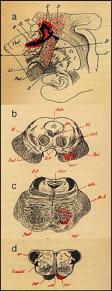
The white matter of the spinal cord contain ascending and descending (motor) fibre pathways. Each of these pathways carry a particular type of impulse. The pathway, which is of particular significance to musle control is the CORTICOSPINAL TRACT. It runs from the motor area of the cerebral cortex to the spinal cord and crosses over at about the lower part of the brain stem.
Motor neurons that synapse above this level are called as UPPER MOTOR NEURONS and those that synapse at or below the level of the anterior horn cells are called LOWER MOTOR NEURONS. If an injury/lesion occur between the brain and the spinal cord i.e proximal to anterior horn, it will be called or considered as an UPPER MOTOR NEURON LESION. Whereas if an injury or lesion occurs between the anterior horn of the spinal cord and the peripheral part, it will be considered as a LOWER MOTOR NEURON LESION.
Read more about UMN on NCBI Bookshelf
Examples of upper motor neuron disease are spinal cord injuries, multiple sclerosis, parkinsonism, CVA etc. Examples of lower motor neuron disease are muscular dystrophies, poliomyelitis, myasthenia gravis and peripheral nerve injuries. In either case of lower motor neuron or upper motor neuron lesion, paralysis usually results, however, the clinical signs differ greatly.
Similar Pages:
Multiple Sclerosis Rehabilitation
Carpal Tunnel Syndrome Exercises
Return from Upper Motor Neuron Lesion to Home Page
Return from Upper Motor Neuron Lesion to Neuro Rehab
Recent Articles
|
Author's Pick
Rating: 4.4 Votes: 252 |

