Enhance your health with free online physiotherapy exercise lessons and videos about various disease and health condition
Sinus Tarsi Syndrome
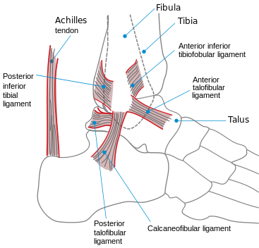
Sinus tarsi syndrome is described as persistent pain at the sinus tarsi that follows a lateral ankle sprain. It has been noted as a sequel of subtalar instability. In addition to pain over the sinus tarsi, patients with this syndrome complain of lateral ankle instability. The cause of the condition is poorly understood and few diagnostic criteria exist. The condition is supposed to be related to fat pad scarring.

Parisien and Vangsness described the use of subtalar joint arthroscopy for evaluation of the posterior facet and its anterior recess. Parisien used this technique to rule out intra-articular disease, to perform biopsy of synovium, and to release periarticular adhesions in three patients with post-traumatic hindfoot pain.
Clinical Signs
- History of foot or ankle inversion sprain- Yes
- Subjective instability- Yes
- Tenderness- Localized to the sinus tarsi
- Ankle stability- Yes/no
- Anesthetic injection to the sinus tarsi- Temporary symptomatic improvement
- Radiographic findings- Normal
- Arthrography- (subtalar) Loss of sinus tarsi filling
- Magnetic resonance imaging- Rupture of interosseous ligament or cervical ligament; bone edema, soft tissue swelling, or fibrosis at sinus tarsi
- Laboratory testing- Rule out systemic inflammatory process
Diagnosis
A diagnostic approach to chronic sinus tarsi pain includes routine and stress radiographs to rule out ankle and subtalar instability as well as MRI to rule out contiguous arthrosis and ganglion formation.
Treatment of Sinus Tarsi Syndrome
- Local injection of anesthetic and corticosteroid is most important for diagnosis and initial empirical treatment.
- Ankle bracing and foot and ankle rehabilitation are used as adjunctive treatments.
- Occasionally, sinus tarsi exploration and debridement are performed either by open method or arthroscopically.
- When indicated, open debridement begins with an incision over the sinus tarsi. Care is taken to avoid the lateral branch of the superficial peroneal nerve. The inferior extensor retinaculum is reflected distally along with the extensor digitorum brevis muscle. The capsule of the subtalar joint is incised. The joint is inspected for chondral injury and osteophytes, which are addressed as needed. Fibrofatty tissue is resected from the sinus tarsi, but the ligaments are kept intact. The wound is closed in a layered fashion with attention to reattachment of the extensor digitorum brevis.
- Patients are placed in a splint postoperatively. After 10 days, the patient is placed into a weight-bearing short-leg cast for 4 more weeks.
- A comprehensive rehabilitation program follows, with an emphasis placed on reducing swelling and improving range of motion, strength, and proprioception.
Return-to-Play
Read Research articles about Sinus Tarsi on PubMed
- Recovery follows a logical sequence of events.
- Once the initial pain and swelling subside, coordination and strengthening activities are emphasized.
- Gradually, the patient is able to return to walking, running, and cutting programs.
- A semirigid pneumatic brace or taping accelerates the schedule.
- Patients are returned to sport once they have mastered sport-specific drills.
Return from Sinus Tarsi Syndrome to Home Page
Return from Sinus Tarsi Syndrome to Sports Physical Therapy
Recent Articles
|
Author's Pick
Rating: 4.4 Votes: 252 |

