Enhance your health with free online physiotherapy exercise lessons and videos about various disease and health condition
Decubitus Ulcer Care

Decubitus Ulcer Care strategies include recognizing risk, decreasing the effects of pressure, assessing nutritional status, avoiding excessive bed rest, and preserving the integrity of the skin. Treatment principles include assessing the severity of the wound; reducing pressure, friction, and shear forces; optimizing wound care; removing necrotic debris; managing bacterial contamination; and correcting nutritional deficits.
A decubitus ulcer is also known as a pressure sore or bed sore and is caused by a lack of blood supply to skin and tissue. Patients who are confined to a bed or chair and need help to move are at greater risk of developing these sores. Pressure sores can be prevented by changing positions frequently, relieving pressure points with extra cushioning, keeping the skin clean, and avoiding dry or chapped skin.
Common causes for Decubitus Ulcer
Shearing and Friction: If a bedridden person is pulled or dragged from his or her bed it causes friction and stretches the skin muscles. Blood circulation of the skin gets marred which causes the damage.
Moisture: Skin is very sensitive at this stage. Perspiration, bed-wetting or feces leads to furthermore chances of bed sores.
Lack of Movement: People, who have been bedridden for a prolonged period of time due to severe medical conditions, bear the brunt. Being in a same position without any movement is one of the main reasons for bed sores.
Lack of Nutrition: A good diet can help you fight this condition easily. Due to lack of proteins, vitamins and other required substances in the body, the patient suffers moreover.
Age: An elderly person is mainly affected because youth is not on his side. The thin skin and failing bodily functions deteriorates the chances of revival.
Lack of Sensation: An injury which leaves you without sensation is another reason for bed sores. This lack of sensation does not allow you to determine the immensity of the pressure applied on the skin.
Decubitus Ulcer Care should focus on these Common sites
About 95% of Decubitus Ulcers occur in the lower part of the body. The areas over the sacrum, coccyx, ischial tuberosities, and greater trochanters account for most Decubitus Ulcer sites. The sacrum is the most frequent site (36% of ulcers), followed by the heel (30%); other body areas like occiput, acromian process, scapula, elbow, lateral and medial malleolus each account for about 6%.
Decubitus Ulcer stages
The definitions of the four decubitus ulcer stages are revised periodically by the National Pressure Ulcer Advisory Panel (NPUAP) in the United States. Briefly, however, they are as follows:
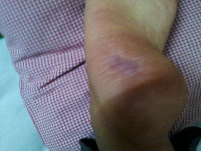
- Stage I is the most superficial, indicated by non blanchable redness that does not subside after pressure is relieved. This stage is visually similar to reactive hyperemia seen in skin after prolonged application of pressure. Stage I pressure ulcers can be distinguished from reactive hyperemia in two ways: a) reactive hyperemia resolves itself within 3/4 of the time pressure was applied, and b) reactive hyperemia blanches when pressure is applied, whereas a Stage I pressure ulcer does not. The skin may be hotter or cooler than normal, have an odd texture, or perhaps be painful to the patient. Although easy to identify on a light-skinned patient, ulcers on darker-skinned individuals may show up as shades of purple or blue in comparison to lighter skin tones.
- Stage II is damage to the epidermis extending into, but no deeper than, the dermis. In this stage, the ulcer may be referred to as a blister or abrasion.
- Stage III involves the full thickness of the skin and may extend into the subcutaneous tissue layer. This layer has a relatively poor blood supply and can be difficult to heal. At this stage, there may be undermining damage that makes the wound much larger than it may seem on the surface.
- Stage IV is the deepest, extending into the muscle, tendon or even bone.
- Unstageable pressure ulcers are covered with dead cells, or eschar and wound exudate, so the depth cannot be determined.
Clinical Presentations which require Decubitus Ulcer Care
The severity of Decubitus Ulceration can be estimated by observing clinical signs. A progression from least tissue damage to most severe damage is presented.
- The first clinical sign of Decubitus Ulceration is blanchable erythema along with increased skin temperature. If pressure is relieved, tissue may recover in 24 hours.
- Progression to superficial abrasion, blister, or a shallow crater indicates involvement of the dermis.
- When full-thickness skin loss is apparent, the ulcer appears as a deep crater. Bleeding is minimal, and tissues are indurated and warm. Eschar formation marks the full-thickness skin loss. Tunneling or undermining is often present.
- The majority of all Decubitus Ulcers develop over six primary bony areas: sacrum, coccyx, greater trochanter, ischial tuberosity, calcaneus (heel), and lateral malleolus.
History need for proper Decubitus Ulcer Care
If an individual has a history of a period of immobility followed by the discovery of a warm, red, spot over a bony prominence , a Decubitus Ulcer can usually be confirmed. If the spot is unnaturally soft to the touch, sometimes reffered to as "boggy", this is enough evidence to suspect that damage is deeper than the epidermis.
Essentials of Decubitus Ulcer Care
Risk Assessment
1. Consider all bed- or chair-bound persons, or those whose ability to reposition is impaired, to be at risk for pressure ulcers.
2. Select and use a method of risk assessment, such as the Norton Scale or the Braden Scale, that ensures systematic evaluation of individual risk factors.
3. Assess all at-risk patients at the time of admission to health care facilities and at regular intervals thereafter.
4. Identify all individual risk factors (decreased mental status, moisture, incontinence, nutritional deficits) to directspecific preventive treatments. Modify care according to the individual factors for proper Decubitus Ulcer Care.
Nutritional status
Malnutrition is frequently cited as a risk factor for the presence, development and non-healing of Decubitus Ulcers. Nutritional status influences the integrity of the skin and support structures, and a lack of vitamins and trace elements may predispose the patient to increased risk of pressure damage. Emaciated and obese people have also been associated with being at a higher risk.
Best practice entails monitoring the nutritional status of individuals as part of a holistic assessment procedure and as an ongoing process throughout an individual’s episode of care. Initially, this assessment should include documentation and monitoring of the following factors for proper Decubitus Ulcer Care:
- current weight and height
- recent weight loss
- usual eating habits
- recent changes in eating habits and intake.
If nutritional risk is suspected, practitioners should undertake more detailed screening.A formal nutritional risk assessment scale may be preferred to help with this and nutritionally compromised individuals should be referred to a dietitian.
Skin Care and Early Treatment
1. Inspect the skin at least daily, and document assessment results for proper Decubitus Ulcer Care.
2. Individualize bathing frequency. Use a mild cleansing agent. Avoid hot water and excessive friction.
3. Assess and treat incontinence. When incontinence cannot be controlled, cleanse skin at time of soiling, use a topical moisture barrier, and select underpads or briefs that are absorbent and provide a quick drying surface to the skin.
4. Use moisturizers for dry skin. Minimize environmental factors leading to dry skin such as low humidity and cold air.
5. Avoid massage over bony prominences.
6. Use proper positioning, transferring, and turning techniques to minimize skin injury due to friction and shear forces.
7. Use dry lubricants (cornstarch) or protective coverings to reduce friction injury.
8. Identify and correct factors compromising protein/ calorie intake and consider nutritional supplementation/ support for nutritionally compromised persons.
9. Institute a rehabilitation program to maintain or improve mobility/activity status.
10. Monitor and document interventions and outcomes for proper Decubitus Ulcer Care.
Mechanical Loading and Support Surfaces
1. Reposition bed-bound persons at least every 2 hours, chair-bound persons every hour.
2. Use a written repositioning schedule for proper Decubitus Ulcer Care.
3. Place at-risk persons on a pressure-reducing mattress/ chair cushion. Do not use donut-type devices.
4. Consider postural alignment, distribution of weight, balance and stability, and pressure relief when positioning persons in chairs or wheelchairs.
5. Teach chair-bound persons, who are able, to shift weight every 15 minutes.
6. Use lifting devices (e.g., trapeze or bed linen) to move rather than drag persons during transfers and position changes.
7. Use pillows or foam wedges to keep boney prominences such as knees and ankles from direct contact with each other.
8. Use devices that totally relieve pressure on the heels (e.g., place pillows under the calf to raise the heels off thebed).
9. Avoid positioning directly on the trochanter when using the side-lying position (use the 30° lateral inclined position).
10. Elevate the head of the bed as little (maximum 30° angle) and for as short a time as possible.
Education
1. Implement educational programs for the prevention of Decubitus Ulcers that are structured, organized, comprehensive,and directed at all levels of health care providers, patients, family, and caregivers.
2. Include information on:
- a. etiology of and risk factors for Decubitus Ulcers,
- b. risk assessment tools and their application,
- c. skin assessment for Decubitus Ulcer Care,
- d. selection/use of support surfaces,
- e. development/implementation of individualized programs of skin care,
- f. demonstration of positioning to decrease risk of tissue breakdown, and
- g. accurate documentation of pertinent data.
3. Include built-in mechanisms to evaluate program effectiveness in preventing Decubitus Ulcers.
Decubitus Ulcer Care should be immediately administered if the formation of pressure ulcers becomes evident.
Further Reading
- Pressure Ulcers and Wound Care. Medscape
- Wound and Pressure Ulcer Management. Johns Hopkins Medicine
- Pressure Sores - Treatment Overview. WebMD
- Bedsores (Decubitus Ulcers). Drugs.com
- Pressure ulcer. A.D.A.M. Medical Encyclopedia. PubMed
- Reddy M, Gill SS, Rochon PA. Preventing pressure ulcers: a systematic review. JAMA. 2006 Aug 23;296(8):974-84.
- Parish LC, Witkowski JA. Controversies about the decubitus ulcer. Dermatol Clin. 2004 Jan;22(1):87-91.
- Bansal C, Scott R, Stewart D, Cockerell CJ. Decubitus ulcers: a review of the literature. Int J Dermatol. 2005 Oct;44(10):805-10.
- Pressure ulcer. From Wikipedia
- Pressure ulcer. MedlinePlus
Return from Decubitus Ulcer Care to Skin Disorders
Return from Decubitus Ulcer Care to Home Page
Recent Articles
|
Author's Pick
Rating: 4.4 Votes: 252 |

